
Ganglion Cysts
A ganglion is a soft tissue mass that most commonly occurs on the wrist in women between 25 and 45 years of age. They are also seen commonly on the foot. A ganglion is a firm, rubbery mass that occurs on the top of the foot. On the foot, the most common area of involvement is in front of the ankle or on the outside of the ankle. A common characteristic of a ganglion is that they will enlarge and then shrink is size. They generally occur without any apparent cause. Ganglions arise spontaneously from a weakness in the soft tissue covering of a joint or tendon sheath. Ballooning out of the tissue occurs and it fills with a thick mucoid fluid. In many instances, ganglions are not painful until they reach a size that causes irritation from shoe pressure. On occasion they will compress a nearby skin nerve and cause tingling into the top of the toes. Tapping on the ganglion will often result in this same tingling sensation into the toes. Other common masses on the foot are giant cell tumors, fibromas and lipomas.
Diagnosis
The diagnosis is made by taking a thorough history of the clinical course of the condition. Physical exam will reveal a firm, rubbery mass that appears encapsulated and will have a discreet boundary. They tend to be firmly adhered to the underlying deep tissues under the skin. A x-ray will reveal the shadow of the soft tissue swelling. On occasion there may be a small bone spur in the area of the ganglion. Spurring indicates a level of arthritis in the joint near the ganglion. A MRI or CT scan will clearly define the mass but is not necessary to make the diagnosis. If a ganglion were suspected within the deep structures of the foot a MRI would be useful to identify the size and extent of the mass.
Treatment
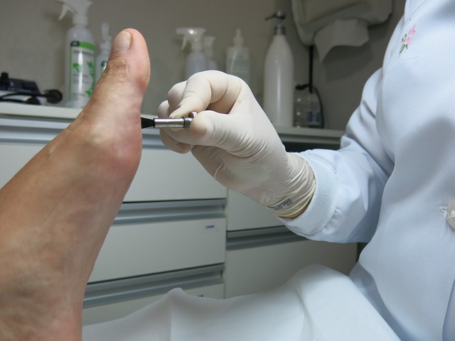
Small ganglions that are not symptomatic or painful usually require no treatment. A non-surgical form of treatment is termed “needling”. This involves numbing the area with a local anesthesia. Once the area is numb a large gauge needle is placed into the ganglion. Aspiration of ganglion fluid is attempted, however, because of the thickness of the fluid it is often difficult to draw the fluid out. The ganglion is then punctured with the needle several times. A steroid medication may then be placed into the mass and a snug bandage applied. This treatment has a 70% recurrence rate. The definitive treatment for a ganglion is surgical excision. (See surgical excision of a ganglion)
Article provided by PodiatryNetwork.com.
Ganglions
A ganglion is a soft tissue mass that most commonly occurs on the wrist in women between 25 and 45 years of age. They are also seen commonly on the foot. A ganglion is a firm, rubbery mass that occurs on the top of the foot. On the foot, the most common area of involvement is in front of the ankle or on the outside of the ankle. A common characteristic of a ganglion is that they will enlarge and then shrink is size. They generally occur without any apparent cause. Ganglions arise spontaneously from a weakness in the soft tissue covering of a joint or tendon sheath. Ballooning out of the tissue occurs and it fills with a thick mucoid fluid. In many instances, ganglions are not painful until they reach a size that causes irritation from shoe pressure. On occasion they will compress a nearby skin nerve and cause tingling into the top of the toes. Tapping on the ganglion will often result in this same tingling sensation into the toes. Other common masses on the foot are giant cell tumors, fibromas and lipomas.
Diagnosis
The diagnosis is made by taking a through history of the clinical course of the condition. Physical exam will reveal a firm, rubbery mass that appears encapsulated and have a discreet boundary. They tend to be firmly adhered to the underlying deep tissues under the skin. A x-ray will reveal the shadow of the soft tissue swelling. On occasion there may be a small bone spur in the area of the ganglion. Spurring indicates a level of arthritis in the joint near the ganglion. A MRI or CT scan will clearly define the mass but is not necessary to make the diagnosis. If a ganglion were suspected within the deep structures of the foot a MRI would be useful to identify the size and extent of the mass.
Treatment
Small ganglions that are not symptomatic or painful usually require no treatment. A non-surgical form of treatment is termed “needling”. This involves numbing the area with a local anesthesia. Once the area is numb a large gauge needle is placed into the ganglion. Aspiration of ganglion fluid is attempted, however, because of the thickness of the fluid it is often difficult to draw the fluid out. The ganglion is then punctured with the needle several times. A steroid medication may then be placed into the mass and a snug bandage applied. This treatment has a 70% recurrence rate. The definitive treatment for a ganglion is surgical excision. (See surgical excision of a ganglion)
Article provided by PodiatryNetwork.com.
Giant Cell Tumor
This tumor was once thought to be a cancer of a tendon sheath. It is now known to be a benign non-cancerous tumor of a tendon sheath. These masses are generally found on the toes, top of the foot or sides of the foot. They are always closely associated with a tendon sheath. They can also occur deep inside the foot. They slowly enlarge but never grow any larger than 4cm in size. They are firm irregular masses that are commonly painful. The pain seems to be a result of the tumor pressing firmly on the surrounding tissues and due to the interference with the function of the tendon that the mass is growing from. As the tendon grows it can press so firmly on the bone it lays next to, that it can cause erosion of the bone. It is because of this erosion of bone that the tumor was once thought to be cancerous. Cancerous tumors can have the characteristic of invading bone through aggressive and destructive means. The erosion of the bone associated with giant cell tumors is due to pressure on the bone and not due to the invasion of the bone by the tumor. Other common soft tissues masses that may occur in the foot are ganglions, fibromas.
Diagnosis
The diagnosis of a giant cell tumor is generally made by a pathologist following removal of the mass. Clinical history of the mass may give the surgeon an idea of what they might expect when removing the mass. X-rays may show the shadow of the mass, and in 10-20% of the cases, may demonstrate bone erosion. The mass is firm and nodular, and always connected to a tendon. A MRI may be useful in determining the extent or size of the mass.
Treatment
Treatment of giant cell tumors is the excision of the tumor. Some physicians may attempt to inject the mass with cortisone in an attempt to shrink the mass.
The Procedure
The surgical excision of giant cell tumors is generally performed in an out patient surgery center. Depending on the location of the mass the surgery may be performed under a local anesthesia, with intravenous sedation or general anesthesia. Following administration of the anesthesia an incision is placed over the mass. The mass is then carefully dissected free from the surrounding soft tissues. Following the closure of the surgical site a gauze compressive dressing is applied. Depending upon the location of the mass the surgeon may apply a splint or below the knee cast. In some instances the surgeon may perfer that the patient use crutches for a few days or for as long as three weeks.
Recovery Period
The recovery period depends upon the location of the mass and the extent of the soft tissue dissection necessary to remove the mass. The sutures are left in place for 10 to 14 days. During this period of time the patient should limit their activities and keep the foot elevated above their heart. It is also important to keep the bandage in place and keep the surgical site dry. If the patient has been instructed to wear a removable cast or use crutches it is important that they follow the surgeons instructions. Time off from work will depend upon the level of activity required of the job and the shoes necessary for work. Generally a minimum of one week off from work is necessary. If the patient can return to work while wearing a cast and they are allowed to perform light duty then they may be able to return to work after one week.
Possible Complications
The surgery is generally successful and without complications. However, as with any surgical procedure there are potential complications. Possible complications include, infection, excessive swelling, delays in healing, tendon or nerve injury. Because the mass is a growth from a tendon, removal of the mass may require the excision of a portion of healthy tendon. This can weaken the tendon or cause scaring of the tendon. Additionally there may be small skin nerves in the area of the tumor that may have to be sacrificed when removing the mass. If this occurs there may be small areas of patchy numbness on the skin following the procedure. This is generally not a significant problem. On occasion a nerve may get bound down in scar tissue and cause pain following the surgery. Recurrence of the mass is also possible but generally not considered a complication of the procedure.
Article provided by PodiatryNetwork.com.
Muco-Cutaneous Cyst
A small nodular single mass that can form on the top of the toe is often times a Muco-Cutaneious Cyst. These occur most frequently at the joint just behind the toenail. These are caused by a weakening of the joint capsule, which allows a swelling to occur. They are firm and rubbery to the touch. Sometimes as the skin thins due to the stretching pressure of the mass it will appear translucent. When the mass is broken or punctured, a thick clear fluid will leak out. If the mass does break open, the area should be kept clean and free of infection. Once the skin heals the mass will reappear.
Treatment
Treatment consists of surgical excision. This can be performed in the doctor’s office under a local anesthesia or in an out patient surgery center. The procedure is relatively simple but can pose a problem for the surgeon, as closure of the skin following removal of the mass can be difficult. Often the surgeon will have to create a skin flap to rotate over the hole where the mass was removed. This requires a bit more of an incision than most patients expect. The foot is bandaged in a dry sterile dressing and the sutures remain in place from 7 to 10 days. The area must be kept dry during this period of time and a limitation of activity is advised. Complications associated with the surgery are infection, delays in healing associated with difficulty in surgically closing the wound. Draining the mass as a form of treatment is not advised unless the patient is made aware of the likely recurrence. Picking the area open at home or attempting to drain it at home is discouraged. An infection in the area could cause permanent joint damage or bone infection.
Article provided by PodiatryNetwork.com.
Plantar Fibromatosis
Within the arch of the foot, firm, nodular masses may form. These can occur as a single mass or in clusters. They are called plantar fibromas and are a non-cancerous tumor that forms within a ligament in the arch of the foot called the plantar fascia. Frequently, they will slowly enlarge causing pain while walking. Their cause cannot always be determined. Damage to the ligament will cause their occurrence and there is an association with taking the drug Dilantin. In 10% of the cases, patients will also demonstrate similar lumps in the palms of the hands called Dupuytren’s Contracture.
Diagnosis
Diagnosis is made by clinical exam. Biopsy of the masses is not recommended. The act of biopsy may cause the fibroma to enlarge. When the mass is removed a definitive diagnosis is provided by examination by microscopic examination by a pathologist
Treatment
Treatment consists of padding the area to reduce pressure. Functional foot orthotics will take the strain off of the plantar fascia ligament and sometimes cause the fibromas to shrink in size. Surgical excision of the mass requires removal of most of the plantar fascia. Simple excision of the mass without removal of the entire ligament generally results in recurrence of the mass. Whenever surgery is contemplated, the patient should wear a functional foot orthotic following the surgery. The orthotic helps to accommodate for the loss of the plantar fascia and its effect on foot function. (See surgical excision of plantar fibromas)
Article provided by PodiatryNetwork.com.
Xanthomas of the Achilles Tendon
Brief Description
An uncommon cause of small lumps in the Achilles tendon is an excessively high cholesterol level in the blood stream. This is a hereditary disorder that results in the deposition of cholesterol in the Achilles tendon. Frequently people will also have yellowish plagues on the lower eye lids, This is a serious condition and requires aggressive treatment by a physician to lower the cholesterol levels. Left untreated the high cholesterol levels can lead to premature heart attack and death.
Diagnosis
Diagnosis is made by clinical exam. Palpation of the Achilles tendon will reveal multiple small nodular masses. Noting excessively high blood cholesterol levels on routine lab tests provides confirmation of the condition. A biopsy of the lesion will also make the diagnosis.
Treatment
The nodules in the Achilles tendon should be left alone. There is no value in removing them. Treatment should be directed at lowering the blood cholesterol levels.
Article provided by PodiatryNetwork.com.
| DISCLAIMER: MATERIAL ON THIS SITE IS BEING PROVIDED FOR EDUCATIONAL AND INFORMATION PURPOSES AND IS NOT MEANT TO REPLACE THE DIAGNOSIS OR CARE PROVIDED BY YOUR OWN MEDICAL PROFESSIONAL. This information should not be used for diagnosing or treating a health problem or disease or prescribing any medication. Visit a health care professional to proceed with any treatment for a health problem. |